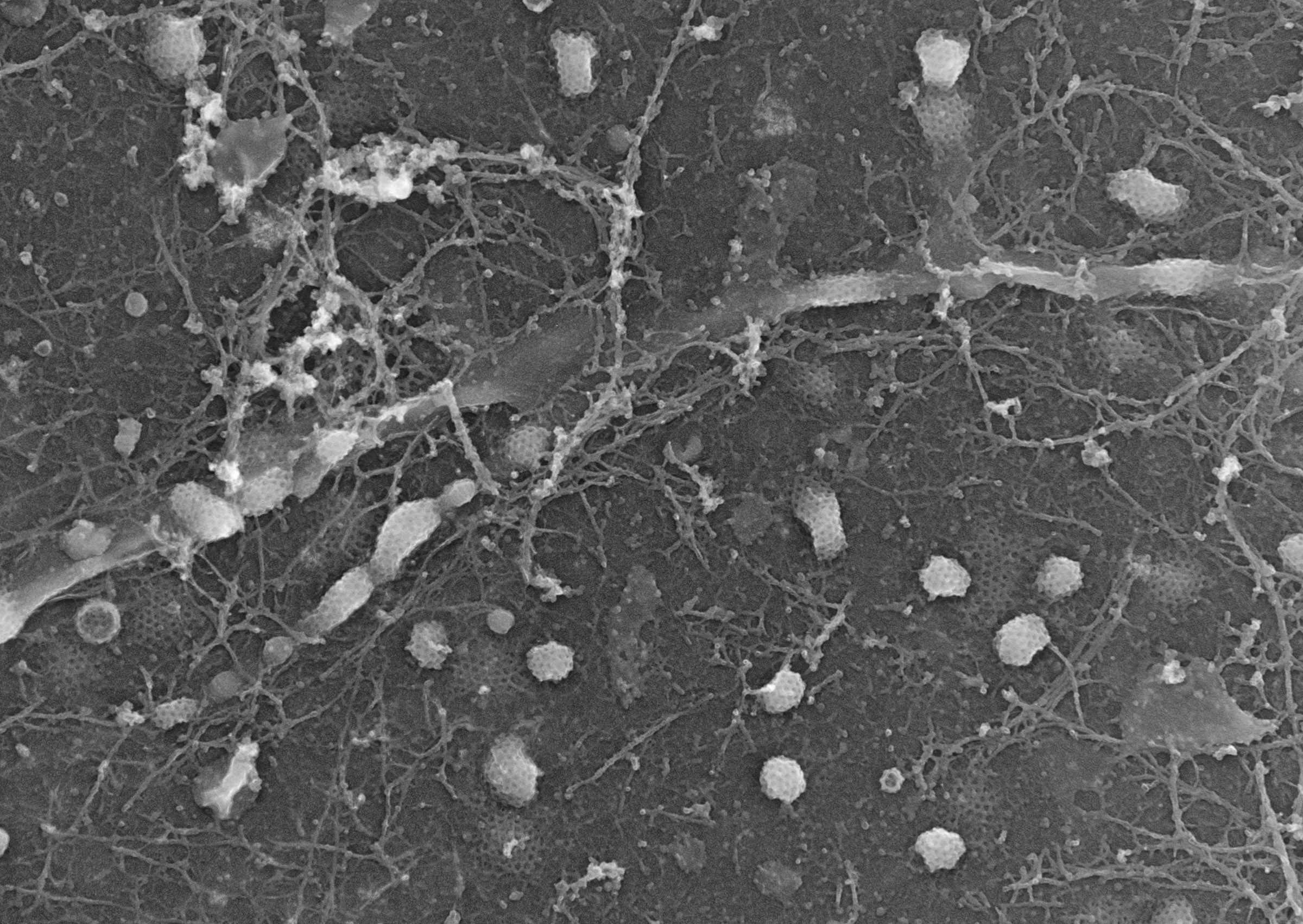
Metal replica electron microscopy of the cell surface
Gallery
Front page of Science with our paper on axonal endocytosis
Cover of NSMB september 2024 issue
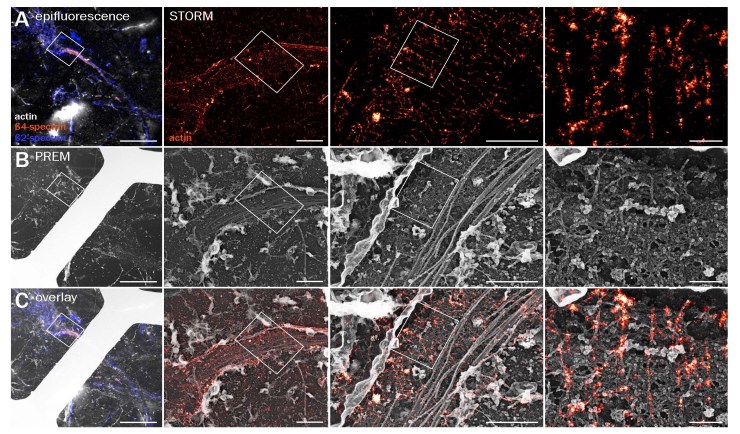
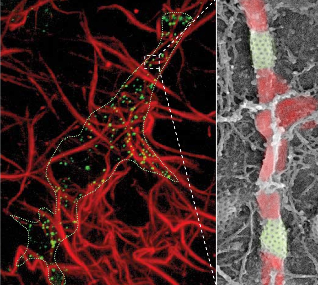
Platinum-replica electron microscopy showing the inner surface of the plasma membrane of a cultured Hs578T cancer cell. Caveolae are pseudocolored purple. Image courtesy of Stephane Vassilopoulos. See Parton et al. Traffic 2020; 21(1): 181–185. Actin rings or braids observed by correlative STORM and platinum-replica electron microscopy. Preprint @bioRxivPlatinum-replica transmission electron microscopy of myotube plasma membranes showing large clathrin lattices (pseudocolored in red) associated with cytoskeletal filaments. We report that these clathrin plaques help to organize desmin intermediate filaments (pseudocolored in purple) and anchor them to the muscle cell membrane. Metal-replica electron microscopy image showing the formation of finger-like actin-based protrusions at the basal membrane of fusing satellite cells. Image is a high-resolution view of the cytoplasmic surface of the plasma membrane from control muscle cells differentiated for 24 h. Protrusions traceable below the acceptor cell are pseudocolored. Quick-freeze, deep-etch transmission electron microscopy of myotube plasma membranes shows an abundance of large clathrin lattices (depicted in various pseudocolors) associated with branched actin filaments. We revealed that these clathrin plaques help to organize skeletal muscle sarcomeres and attach them to the muscle cell membrane. Metal-replica electron microscopy image showing the formation of clathrin tubes pinching collagen fibers on the cytoplasmic side of the membrane from MD-231 cells. Collagen fibers are pseudo-colored in red on the EM image. (Elkhatib et al., 2017).